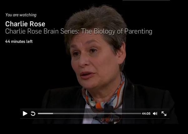
Scientific American Features Story by Professor Hensch:
The Power of the Infant Brain:
What’s on your music player? If you’re older than 30 years, it probably includes songs from your teenage years. Childhood and adolescence are the most impressionable period of a person’s life. The earliest memories and experiences are essential in shaping character—and they profoundly influence everything that comes next. “The habits we form from childhood make no small difference, but rather they make all the difference,” Aristotle proclaimed more than 2,000 years ago.
Read the full article by Conte Center director Professor Takao Hensch in Scientific American >>
Related Medscape Interview with Prof. Hensch:
Harnessing the Childhood Brain to Treat Alzheimer Disease, Autism, and Mental Illness >>
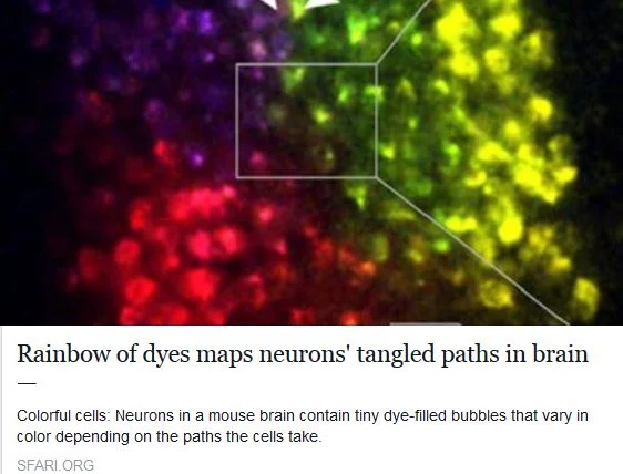
Imaging Neural Connections:
New study raises the bar for precision, speed and thoroughness
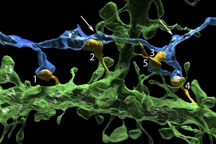
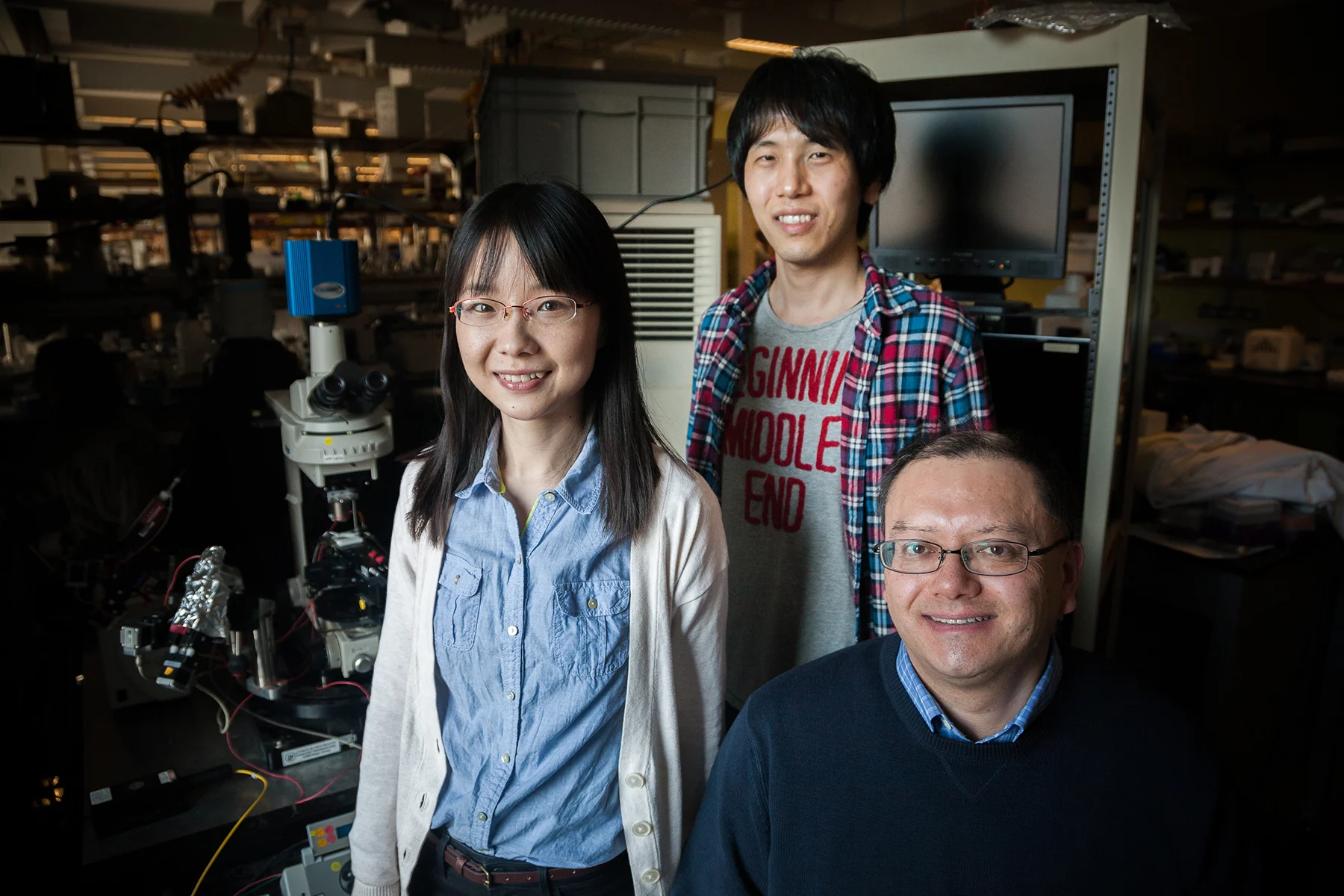
Team from Zhuang lab makes first super-resolution microscopy map of all the inhibitory synapses in a neuron’s dendritic tree

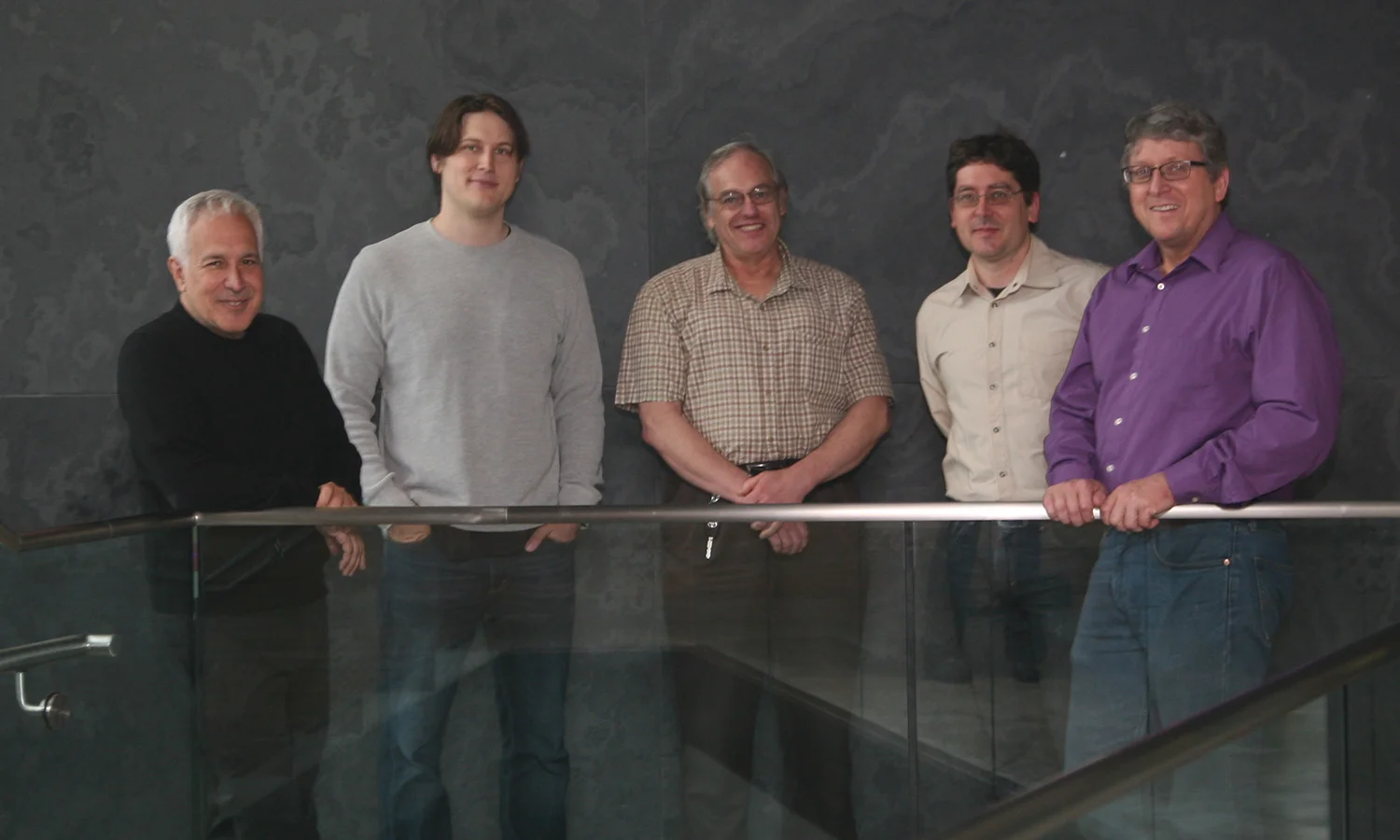
Yaron Sigal, Colenso Speer, Hazen Babcock and Xiaowei Zhuang
(Photograph courtesy of Kendal Kelly)
Geographical maps have long changed the way we live, from the ancient hand-drawn ones that relied on explorer voyages to those on today’s satellite-driven, real time apps like Google Maps. The same transformative power holds true for maps in neuroscience. From the meticulous illustrations of neurons in different brain regions made by neuroanatomist Ramon y Cajal in the late 1800s to the imaging of neural circuits through more modern computerized technologies, the maps that we have of our nervous systems are constantly shaping the questions we think to ask and are able to answer about how our brains and bodies work—and what to do when diseases or injuries arise.
A recent advance in the map-making world of brain scientists comes from the lab of Xiaowei Zhuang, a faculty member in the Conte Center at Harvard who is the David B. Arnold Professor of Science and a Howard Hughes Medical Institute Investigator. In a study published in Cell, led by postdoctoral fellows Yaron Sigal and Colenso Speer, her team showcases a powerful new approach for visualizing and analyzing the input fields of neurons, using neurons in the eye called retinal ganglion cells as a test case. While mapping the sea of inputs that a single neuron receives and integrates is something that neuroscientists have long recognized as critical for understanding the functions of different kinds of neurons and their role in circuits—and have been able to accomplish in varying capacities—until now there’s usually been a trade-off between obtaining very high resolution images and having precise molecular information. The Zhuang team’s new strategy will allow researchers automated analysis of neuron input fields in a way that minimizes this compromise and overlays complex molecular information upon three dimensional reconstructions of neurons situated in relatively thick sections of brain tissue.
Resolution and specificity are factors that matter to map-makers of all kinds. To give a Google Maps analogy, it’s like wanting the power to zoom in deep on any location of interest (say to look for a parking lot near an event venue) and at the same time, the power to selectively tag points of interest within a dense field (for instance, to highlight all the coffee shops along a route). In neuroscience terms, this might mean desiring the ability to zoom in highly upon the input and output structures of individual neurons in the brain, called dendrites and axons. And at the same time, wanting the ability to selectively tag all the synapses—the points of communication between neurons—of a particular variety that dot these dendrites and axons. Read more >>
Imaging the Brain’s Jungles:
A Miniscule Yet Monumental Leap Forward
The Incredible Amount We Can Learn from Electron Microscopy Reconstruction of Less Than 0.000001% of the Mouse Brain
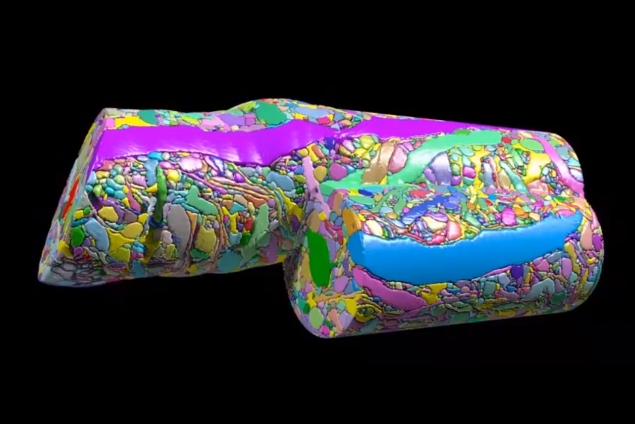
When it comes to science, perhaps the wisest amongst us are those who know how much we don’t know. The famous 19th century neuroanatomist Santiago Ramón y Cajal, who many would argue did more to characterize the different cell types that make up the nervous system than any other scientist of his time, once described the adult brain as a ‘jungle’ that is ‘impenetrable and indefinable.’
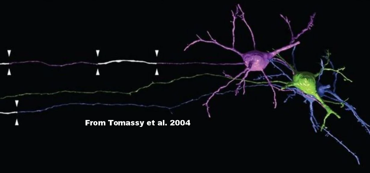
Over a hundred years later, despite an explosion of new technologies in microscopy and genetic and immunological methods of brain cell labeling, neuroscience research remains chock full of uncharted territories. In a recent publication in Cell, a team led by Jeff Lichtman, the Jeremy R. Knowles Professor of Molecular and Cellular Biology at Harvard—fittingly also named the Santiago Ramón y Cajal Professor of Arts and Sciences—presents the latest chapter in this story, by providing unparalleled views into the intricacies of synaptic connectivity in the cerebral cortex and revealing novel insights about this connectivity—while simultaneously highlighting just how much we have left to discover.
The new study offers the first detailed foray into the neocortex, with a stunningly saturated reconstruction of a volume of mouse cortical tissue of about 1,500 cubic microns obtained at a resolution fine enough to allow the identification of every single synaptic vesicle within. This is remarkable considering how incredibly small synaptic vesicles are, how many synaptic vesicles are present at each synapse, and how many different synapses each neuron has. Read more >>
More Stories


From discovery to trial: A drug that may correct ‘lazy eye’
A blog post by Sarah Lewin in Vector, Boston Children’s Hospital’s science and clinical innovation blog